
Why Türkiye Has Become a Reference Point for Skin Surveillance
Skin cancer is one of the few cancers where early detection genuinely changes the outcome. A melanoma found at 0.8 mm carries a very different prognosis from one found at 3 mm, and the difference between those two numbers is usually not treatment — it is surveillance. This is the principle that has quietly turned several Turkish hospital dermatology departments into destinations for international patients who want their skin checked properly, not quickly.
Türkiye's advantage here is structural rather than accidental. The country operates a dense network of university hospitals and large private hospital groups where dermatology, dermatopathology and plastic surgery sit in the same building, often on the same floor. That physical proximity means a suspicious lesion can move from dermatoscopic image to excisional biopsy to microscope review without the patient being sent across a city — or across a border — to find the next specialist.
For medical travellers, the practical consequence is significant. Programme-based mole mapping and dermatopathology in Türkiye are typically organised so that a thorough assessment — history, total-body photography, digital dermatoscopy of every relevant lesion, and where indicated a biopsy with same-week pathology — can be completed within a single visit window of a few days, with images and written reports handed to the patient for ongoing monitoring at home.
What Digital Dermatoscopy Actually Adds to a Skin Check
Digital dermatoscopy (also called dermoscopy or videodermatoscopy) is the use of a specialised magnifying instrument with controlled polarised lighting to examine skin structures below the surface reflection that the naked eye cannot see. A dermatologist using dermatoscopy is not simply looking at a bigger mole; they are reading pigment network patterns, vascular structures, regression areas and distribution of dots and globules that correspond to specific histological features.
In a hospital-based programme, the instrument is linked to imaging software. Each lesion is captured, measured, tagged to a body-map location and stored. On a repeat visit — whether six months or two years later — the software can place the old and new images side by side at identical magnification. Subtle changes in asymmetry, border or colour that would be almost impossible to detect by eye become visible as a direct comparison.
Turkish dermatology centres have invested heavily in this technology, and many use high-resolution digital systems with automated or semi-automated change detection. During your consultation, ask which system is used, whether images are stored long-term in your file, and whether you will receive digital copies. Most hospital-based programmes in Istanbul, Ankara and Izmir will provide these as standard, and the images become far more useful when you can share them with your own dermatologist at home.
Total-Body Mole Mapping: The Full Picture, Not Just the Spot
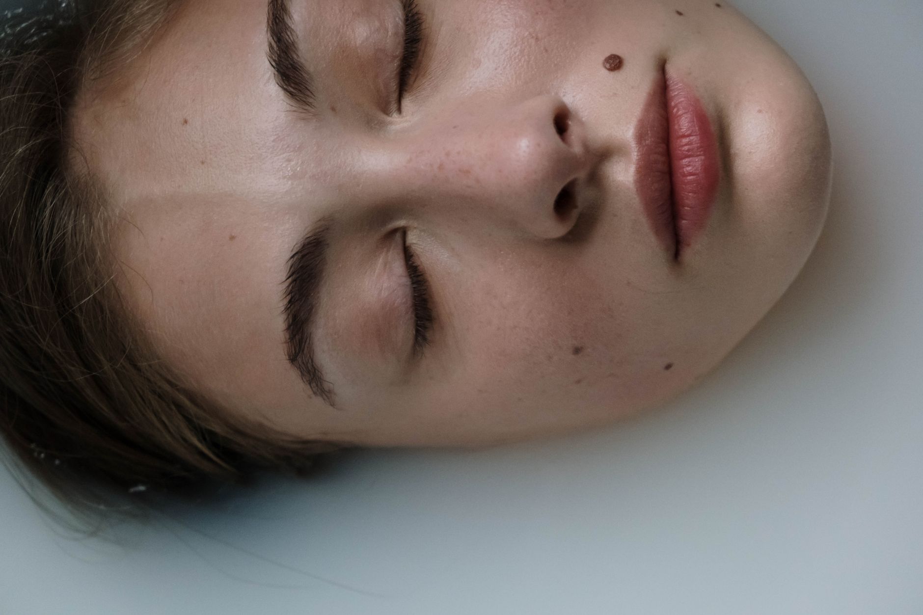
Mole mapping — also called total-body photography or digital follow-up — is the systematic photographic documentation of the entire skin surface. It exists because melanoma surveillance is not only about evaluating one lesion; it is about knowing which lesions a patient has, where they are, and how they behave over time.
A proper mapping session begins with a standardised set of macro photographs covering the body in defined views: front, back, both sides, arms, legs, hands, feet, scalp and, where relevant, mucosal surfaces. Each image is calibrated for distance and lighting so that future sessions are comparable. The dermatologist then examines the skin with dermatoscopy, marks any lesion of interest, and correlates it to the body map.
The value of this is cumulative. A patient with many atypical naevi — sometimes called atypical mole syndrome or dysplastic naevus syndrome — may have dozens of lesions that all look slightly unusual. Individually, each may be benign. The question that matters is whether one of them is changing. Without a baseline map, that question can only be answered by memory; with a map, it is answered by direct comparison.
Hospital-based programmes in Türkiye usually combine total-body photography with dermatoscopy of individual lesions in a single appointment. A typical session takes 45 to 90 minutes depending on the number of lesions and the need for close-up imaging. The output is a structured report: body-map images, dermatoscopic images of flagged lesions, and a recommendation for the next follow-up interval.
Dermatopathology: Where the Diagnosis Is Actually Made
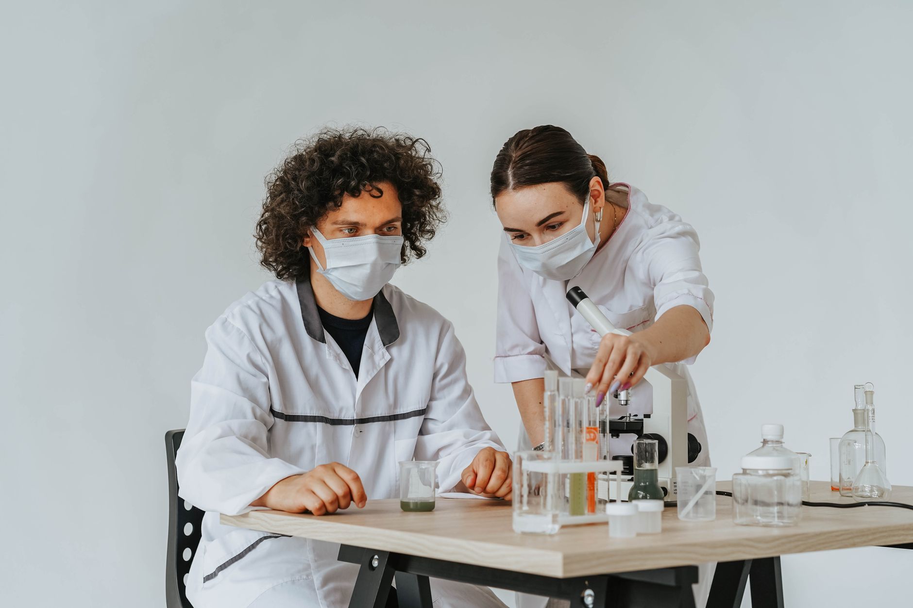
Dermatopathology is the subspecialty that examines skin biopsies under the microscope. It is the reference standard for diagnosing melanoma, atypical naevi, basal cell carcinoma, squamous cell carcinoma and a long list of inflammatory skin diseases. In many health systems, dermatopathologists are scarce, and biopsy results can take weeks to return with limited subspecialty input.
In Türkiye's larger hospitals, dermatopathology is an integrated part of the dermatology department. Biopsies taken in the clinic are processed in-house, and the dermatopathologist works directly with clinical images — including the dermatoscopic photographs taken before the biopsy. This matters because a lesion's dermatoscopic appearance can guide the pathologist's sectioning strategy and interpretation, particularly in borderline lesions where the distinction between a severely atypical naevus and an early melanoma can be genuinely difficult.
Immunohistochemistry is widely available, including markers such as S100, SOX10, Melan-A, HMB-45 and PRAME, which are used to clarify melanocytic lesions. Many centres also offer molecular testing where indicated. For patients travelling to Türkiye specifically for a second opinion on a previously read biopsy, this combination — expert dermatopathology plus the original clinical and dermatoscopic context — is often the single most valuable service available.
How a Single-Visit Assessment Is Structured
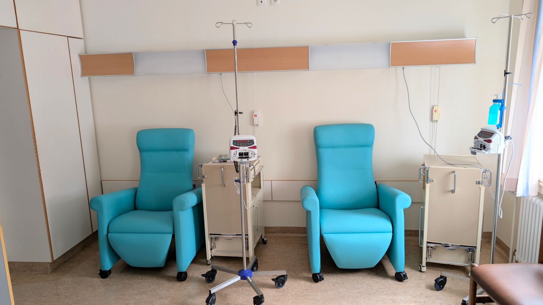
One of the most common questions from international patients is whether everything can realistically be done in one trip. In most hospital-based programmes, the answer is yes for the assessment, and usually yes for minor procedures as well. A typical schedule looks like this.
- Day 1 — Consultation and imaging. Full skin examination, total-body photography, digital dermatoscopy of all relevant lesions, and a documented risk assessment based on personal and family history, skin type and previous sun exposure.
- Day 1 or 2 — Biopsy if needed. Suspicious lesions are removed or sampled under local anaesthesia. Most biopsies take 15 to 30 minutes, and patients leave with a dressing and written aftercare instructions.
- Day 3 to 5 — Pathology review. Specimens are processed and reviewed by the dermatopathologist. Many centres prioritise international patients and can return results within a few working days.
- Day 5 or 6 — Results consultation. The dermatologist explains the findings, provides a written report in English (or your preferred language), and outlines follow-up recommendations.
Not every patient needs a biopsy — in fact, most do not. For patients whose lesions are all stable and benign-appearing, the visit is purely diagnostic and preventive, and the deliverable is a baseline map plus a surveillance plan.
Who Benefits Most from This Kind of Programme
Mole mapping and dermatopathology programmes are not necessary for everyone, but they are disproportionately valuable for specific groups. If you recognise yourself in any of the following categories, a structured assessment is worth considering.
- People with many moles or atypical moles. More than 50 to 100 naevi, or several clinically atypical ones, raises baseline melanoma risk and makes change detection harder without a map.
- Those with a personal or family history of melanoma or other skin cancer. Surveillance intervals should be shorter and the assessment more detailed.
- People who have had significant sun exposure or sunbed use, particularly with episodes of blistering sunburn in childhood or adolescence.
- Immunosuppressed patients, including transplant recipients and those on certain long-term medications, who face elevated skin cancer risk.
- Patients seeking a second opinion on a biopsy that was read elsewhere, or on a lesion that has been described as borderline or atypical.
For these groups, the combination of dermatoscopy, total-body photography and in-house dermatopathology is not excessive — it is the appropriate standard, and it is precisely what hospital-based Turkish programmes are built to deliver.
The Reassuring Precision of Turkish Dermatologists
What international patients consistently comment on is not the technology alone, but how it is used. Turkish dermatologists in these programmes tend to be meticulous in a very specific way: they document everything, they explain their reasoning, and they do not rush the decision to biopsy or the decision not to.
This matters because over-biopsy is a real harm. Removing benign lesions unnecessarily leaves scars, creates anxiety and generates cost. Under-biopsy is a worse harm. The skill lies in knowing which lesions can be safely monitored with imaging and which require tissue. Dermatoscopy, combined with a baseline map and expert pathology backup, is what makes that judgement reliable rather than intuitive.
Patients also value the directness of the reporting. Rather than a vague reassurance that everything looks fine, they receive a written report with images, lesion locations, dermatoscopic descriptions and a recommended follow-up interval. That document is portable. It can be handed to a dermatologist at home, compared against future imaging, and used to justify insurance coverage for ongoing surveillance.
Practical Planning: Costs, Timelines and Follow-Up
Costs for dermatology and dermatopathology in Türkiye are typically well below those in Western Europe or North America, but the more important variable is efficiency. A programme-based assessment that might require three separate appointments in another system is often compressed into one or two visits, which reduces travel time, accommodation costs and time away from work.
When enquiring with a hospital, ask for a written plan that specifies: the type of dermatoscopy system used, whether total-body photography is included, whether images and reports are provided digitally, the expected turnaround time for pathology, and the cost of biopsy and histology as separate line items. Reputable centres will provide this without hesitation.
Follow-up intervals are usually individualised. Low-risk patients with a stable baseline map may be advised to return every 12 to 24 months. Higher-risk patients, or those with a history of melanoma, are often advised every 3 to 6 months, at least initially. The baseline images you take home are the key to making those intervals meaningful — and to ensuring that whatever happens next, your skin is being compared against a documented past rather than a fading memory.
Bringing It Together: A Realistic Expectation
A well-run dermatopathology and mole mapping programme in Türkiye offers something that is surprisingly hard to find elsewhere as a package: comprehensive imaging, expert clinical judgement, same-week histopathology and a portable record of your skin. It is not a miracle and it does not replace ongoing care at home. It is a rigorous, documented baseline — and for many patients, that is exactly what has been missing.
If you are considering travelling for this kind of assessment, the most useful first step is to gather your existing records: previous biopsy reports, photographs of lesions, and any dermatology notes. Send them ahead. A good centre will review them before you arrive, tell you honestly whether a trip is worthwhile, and design a visit that answers the questions you actually have.
Thinking about treatment in Turkey?
Share your contact details and a verified doctor will follow up with a plan built around you — free, no obligation.